How to cite: Gupta GN. Surveillance after focal therapy. Grand Rounds in Urology. Published October 2025. Accessed February 2026. https://grandroundsinurology.com/surveillance-after-focal-therapy/
Summary
Gopal N. Gupta, MD, FACS, Professor of Urology, Radiology, Surgery, Loyola University, Chicago, Illinois, describes principles of surveillance following focal therapy for prostate cancer. He states that although technique and planning continue to advance, surveillance determines long-term success.
He notes that focal therapy carries a higher local failure rate than radical therapy, including residual disease within the treated zone, new infield disease, and out-of-field cancer. Long-term oncologic data remain limited, and predictors of recurrence are still under development. He explains that surveillance relies on prostate-specific antigen (PSA), post-ablation biopsy, and multiparametric magnetic resonance imaging (MRI) with supplemental evaluation by emerging imaging such as prostate-specific membrane antigen positron emission tomography (PSMA PET).
PSA is the most widely used marker, yet it performs poorly for detecting residual disease. He cites data in which PSA nadir, six-month PSA, and PSA density all demonstrate limited predictive value for recurrent clinically significant cancer. He then reviews MRI interpretation after ablation, noting that expected findings vary across T2 diffusion and contrast-based sequences. MRI at six months performs better than very early MRI and offers a more substantial negative predictive value for higher-grade recurrence.
He discusses a small series evaluating PSMA PET after ablation, which detects some recurrences missed by MRI, yet still has false negatives for low-volume significant disease. He introduces an international consensus recommending PSA at three months and every six months thereafter, MRI at six months, and annual imaging and biopsy at twelve months or sooner if indicated.
Dr. Gupta shares his institutional approach, which includes structured PSA schedules, MRI at 12 months, targeted and systematic biopsy, team-based radiology and pathology review, and the integration of PSMA and predictive modeling to refine risk assessment over time.
Frontiers in Oncologic Prostate Care and Ablative Local Therapy (FOCAL) is an outstanding program on prostate imaging, transperineal interventions, and ablative treatments for prostate cancer and benign prostatic hyperplasia. Bringing together community-based, academic, and industry partners, FOCAL offers lectures by world-renowned faculty and hands-on training workshops on in-office transperineal interventions, fusion-guided prostate ablation and state-of-the-art BPH management with novel technologies.
ABOUT THE AUTHOR
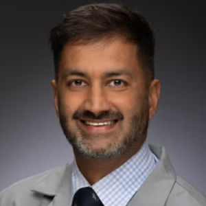
Gopal N. Gupta, MD, FACS, is Professor of Urology, Radiology, and Surgery at Loyola University Chicago Stritch School of Medicine and Director of the Urologic Oncology Fellowship at Loyola Medicine. He is a fellowship-trained urologic oncologist specializing in the diagnosis and treatment of genitourinary malignancies, with particular expertise in prostate, bladder, and kidney cancers. His clinical interests include robotic and minimally invasive surgery, advanced imaging, MRI-ultrasound fusion-guided prostate biopsy, molecular imaging, and focal therapies for prostate cancer.
Dr. Gupta earned his medical degree from the University of Illinois at Chicago College of Medicine and completed his urology residency at the University of Maryland Medical Center. He subsequently completed a fellowship in urologic oncology at the National Cancer Institute, part of the National Institutes of Health, where he trained in the Urologic Oncology Branch under W. Marston Linehan, MD. Dr. Gupta is actively involved in clinical education, research, and the multidisciplinary management of patients with urologic cancers.