Steven S. Raman, MD

Steven S. Raman, MD

University of California, Los Angeles

Los Angeles, California

Steven S. Raman, MD, is the Director of the Abdominal Imaging Fellowship Program, Director of the UCLA Uterine Fibroid Program, and a Clinical Professor of Radiology Sciences at the University of California, Los Angeles. Dr. Raman is also Director of the Abdominal Imaging Fellowship at UCLA, and Co-Director of the Fibroid Treatment Program at UCLA. He is an expert in abdominal and pelvic imaging and interventional radiology, especially in the area of tumor ablation and fibroid treatment. 

Dr. Raman earned his medical degree from Keck School of Medicine in Los Angeles, California. He completed an internship in Internal Medicine at the Pacific Presbyterian Medical Center in San Francisco, California. Dr. Raman then completed a residency in Radiological Sciences at University of California, San Diego. He then completed a fellowship in Abdominal Imaging at the University of California, Los Angeles. 

Dr. Raman has been actively involved in research involving abdominal imaging and cross-sectional interventional radiology. His research interests also include developing and improving techniques in tumor ablation and general interests in liver, pancreas and genitourinary tract imaging. Dr. Raman has 579 publications within his field, and is a member of the Cancer Molecular Imaging, and the Nanotechnology and Theranostics groups within UCLA.

Talks by Steven S. Raman, MD

Integration of Histology, Genomics and Proteomics with MRI

Steven S. Raman, MD, explores how histology, genomics, and proteomics with MRI revolutionize the diagnosis and treatment of prostate cancer. Histology provides critical insights into the pathological features of prostate cancer; genomics offers a deeper understanding of the genetic mutations and alterations that drive cancer progression; and proteomics sheds light on the protein expressions and interactions that occur within the tumor microenvironment. Raman asserts that each modality, while powerful on its own, gains significance when integrated with MRI prostate imaging.

He elaborates on the technological advancements and methodological innovations that facilitate this integration. Raman highlights how multi-parametric MRI (mpMRI) can be augmented with molecular data from histology, genomics, and proteomics to create a more comprehensive and nuanced diagnostic picture. Recent studies show significant improvements in the detection, characterization, and monitoring of prostate cancer, leading to more personalized and effective treatment strategies.

Read More
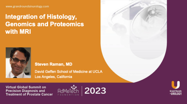
Image-Guided Minimally-Invasive Whole Gland Treatment

Steven S. Raman, MD, Clinical Professor of Radiology and Director of Prostate MR Imaging and Interventions at the David Geffen School of Medicine of the University of California, Los Angeles, explains how magnetic resonance imaging (MRI) aids in prostate cancer staging, diagnosis, follow-up, treatment planning, and image-guided treatment. After reviewing relevant FDA guidelines, Dr. Raman outlines the MRI-guided transurethral ultrasound ablation (TULSA)-PRO® treatment planning and delivery process, noting the potential for real-time MR thermometry and feedback control. To further display benefits of the treatment, he reviews outcomes from the TACT trial. He then addresses potential next steps in prostate tissue ablation.

Read More