Nelson N. Stone, MD, presented “Transperineal Mapping Biopsy Defines Tumor Type and Location” during the 24th Annual Southwest Prostate Cancer Symposium on April 13, 2019 in Scottsdale, Arizona.
How to cite: Stone, Nelson N. “Transperineal Mapping Biopsy Defines Tumor Type and Location” April 13, 2019. Accessed Apr 2026. https://grandroundsinurology.com/transperineal-mapping-biopsy-defines-tumor-type-and-location/
Transperineal Mapping Biopsy Defines Tumor Type and Location – Summary:
Nelson N. Stone, MD, reviews the limitations of systematic biopsy and MRI in regard to accurately localizing and grading of prostate cancer tumors. He describes a 3D transperineal biopsy platform that could improve tumor localization and provide a pathway to targeted focal therapy.
Abstract:
In order to effectively plan for prostate cancer focal therapy, it is essential to precisely locate and grade all lesions in the gland. Currently, 12-core systematic biopsies are unsatisfactory for this purpose.
MRI can be beneficial in terms of identifying targets for re-biopsies in patients with a previous negative or low-risk biopsy but continued clinical suspicion of cancer or higher-risk disease. However, MRI has significant limitations. An analysis of correlation between MRI results and whole-mount histopathology showed that MRI misses 73%-89% of lesions smaller than 10mm in diameter. MRI also underestimates the size and extent of prostate tumors, and has been shown to leave 49% of lesions outside the ablation field when used to plan focal therapy.
A modality that may be able to adequately define a patient’s tumor type and location is transperineal mapping biopsy (TPMB). However, typical template-based TPMB presents challenges related to instrumentation, software, and maintaining the integrity of specimens taken.
This presentation reviews a TPMB platform that could address these challenges. This platform’s needle has the ability to take samples up to 60mm in length, with a trocar tip and an improved cannula design that mitigates sample compression and fragmentation, as well as needle deflection. The biopsy actuator allows for adjustment of the needle in order to take varying specimen lengths depending on the size of the gland.
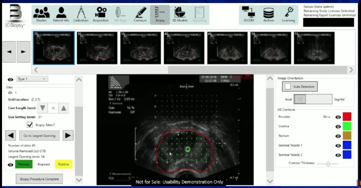
With the platform’s software, physicians can record pathology information and recreate a dimensional display of the length, position, and orientation of each sample and use it to plan ablation zones.
The platform also includes a pathology carrier system to avoid the fragmentation and misalignment of cores that often occurs during specimen handling.
About the Southwest Prostate Cancer Symposium
The Southwest Prostate Cancer Symposium (SPCS) is a multi-day conference that seeks to educate urologists, radiation oncologists, medical oncologists, and other healthcare professionals involved in the treatment of prostate cancer. The topics focus on current technical aspects of diagnosis and treatment of localized and advanced disease, particularly regarding imaging, technology, and training in the related devices. Dr. Stone presented this lecture during the 24th SPCS in 2019. In 2020, the 25th SPCS will also offer training sessions involving imaging, scanning, and prostate cancer treatment- related devices on site. Please visit this page in order to register for future SPCS meetings.
ABOUT THE AUTHOR
Nelson N. Stone, MD, is Professor of Urology, Radiation Oncology, and Oncological Sciences at the Icahn School of Medicine at Mount Sinai and chief medical officer at Viomerse, Inc.
Dr. Stone earned his medical degree from the University of Maryland in 1979. He completed a Residency in General Surgery in 1981 at the University of Maryland, followed by a Residency in Urology at the University of Maryland. He then completed a Fellowship in Urologic Oncology at Memorial Sloan-Kettering Cancer Center and a Research Fellowship in Biochemical Endocrinology at Rockefeller University in 1986. He was Chief of Urology at Elmhurst Hospital Queens from 1986-1996.
Dr. Stone has founded several medical companies and serves on the editorial board of many scientific journals. He is a member of many professional societies, including the Prostate Conditions Education Council, the Society for Minimally Invasive Therapy, the New York State Urological Society, the American Association of Clinical Urologists, and the American Urologic Association. Dr. Stone has participated in approximately 25 research studies on prostate cancer and has authored more than 500 articles, abstracts, and book chapters, primarily on prostate cancer. He invented the real-time technique for prostate brachytherapy in 1990 and has trained more than 5,000 physicians worldwide through his company ProSeed. His most recent company, Viomerse, creates synthetic body parts (phantoms) for surgical training and has recently released an extended reality remote training platform.