Francisco G. La Rosa, MD

Francisco G. La Rosa, MD

University of Colorado, Anschutz Medical Campus

Aurora, Colorado

Francisco G. La Rosa, MD, FCAP, serves as an Associate Professor in the Department of Pathology at the University of Colorado Anschutz Medical Campus and is a distinguished member of the University of Colorado Cancer Center. He originally graduated as an M.D. in Lima, Peru, with Residency in Clinical Pathology, followed by Residency in Anatomic and Clinical Pathology at the “University of Colorado.” He is Board Certified in anatomic and clinical pathology, and specializes in genitourinary, renal, and heart pathology. He is also recognized as an expert in pathology informatics.

Dr. La Rosa actively engages in clinical practice, research, and education with over 100 publications in refereed journals. His primary hobbies include photography and videography, both of which he has utilized for educational purposes by creating several websites dedicated to the teaching of pathology.

Dr. La Rosa holds memberships in several national and international organizations, including the College of American Pathologists, American Urological Association, Peruvian American Medical Association, Catholic Medical Association and Association for Pathology Informatics. His research involvement extends to collaboration on various research grants in genitourinary pathology, and he serves as a member and study coordinator at the Southwest Oncology Group (SWOG), Genitourinary Committee. Most notably, he collaborates closely with Dr. E. David Crawford and his team on cutting-edge technology for diagnosing and treating prostate and urinary cancers. As a faculty member of Grand Rounds in Urology, he actively participates in various innovative educational activities within the field of urology.

As a graduate from the Teaching Scholar Program at the University of Colorado, Dr. La Rosa imparts his knowledge to medical students, as well as pathology residents and fellows. His commitment to excellence in teaching has earned him several recognitions.

Beyond national borders, Dr. La Rosa is recognized as an Honorary Professor at the Medical School Hipolito Unanue of the National University Federico Villarreal in Lima, Peru. He also serves as the co-founder and current Administrator of the Asociación Iberoamericana de Telesalud y Telemedicina (AITT) and holds the position of founder and Editor-in-Chief of the international journal Revista de la AITT (ISSN 2411-3840). The mission of these endeavors is to promote and coordinate programs and activities in telehealth and telemedicine among Latin American countries. Additionally, he serves on several international scientific committees and editorial boards of scientific journals, further contributing to the global advancement of pathology.

Disclosures:

Talks by Francisco G. La Rosa, MD

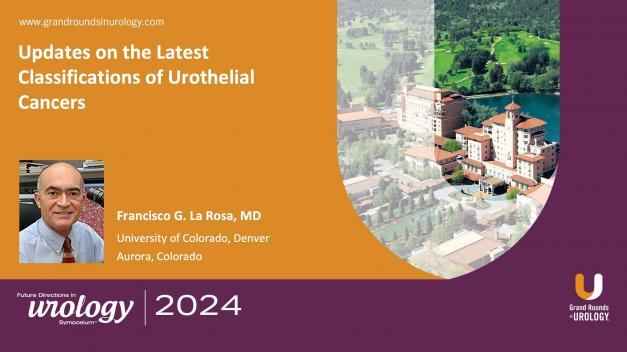
Updates on the Latest Classifications of Urothelial Cancers

Francisco G. La Rosa, MD, provides an overview of the historical evolution and recent updates in cancer classification.

In this 11-minute presentation, he highlights the early stages of pathology in the 1800s. Key advancements emerged with the establishment of histopathology in the early 1900s, marking significant strides in cancer characterization.

La Rosa reviews refinements in classifications, such as the 1952 Armed Forces Institute of Pathology system, the WHO’s 1973 classification, the WHO and International Society of Urological Pathology 1998 collaboration, and, finally, the 2022 WHO update. This final update represents a major advancement by incorporating molecular insights and enhancing cancer classifications’ prognostic and therapeutic implications. This edition also discards ambiguous terms like “urothelial dysplasia” and “papillary proliferation of undetermined malignant potential” to reduce diagnostic variability.

Additionally, the updated classification system includes detailed subtypes based on molecular markers, supporting a more nuanced understanding of tumor behavior. The Cancer Genome Atlas is pivotal in this molecular stratification, identifying specific cancer subtypes and associated prognostic markers.

Read More
Pathological Aspects of BPH, Prostatitis, and Other Painful Co-Occurrences

Francisco G. LaRosa, MD, FCAP, explores the pathology of benign prostatic hyperplasia (BPH), prostatitis, and associated urological conditions. He begins by reviewing the pathophysiology of BPH, presenting examples of usual and specific patterns of BPH and their progression.

Transitioning to prostatitis, Dr. LaRosa examines acute and chronic prostatitis. He highlights the mechanisms behind its close association with BPH, despite its frequent misdiagnosis and mismanagement in BPH patients.

Dr. LaRosa concludes by providing examples of other non-cancerous co-occurrences which can be discovered after prostate biopsy. He focuses on fibromas, thromboembolisms, and calcification.

Read More

The Future of AI in Pathologic Evaluation of Prostate Cancer

Francisco G. LaRosa, MD, FCAP, explores possible applications for artificial intelligence (AI) in prostate cancer detection and treatment. He begins by outlining the potential benefits of AI in prostate cancer evaluation.

Dr. LaRosa then discusses the technological innovations which allowed AI to be incorporated as a tool in prostate cancer detection. He presents current examples of AI being used in prostate cancer screening.

However, Dr. LaRosa concludes by cautioning against relying on AI He outlines current issues with machine-vision capabilities, detection models, unclear objectives, and contaminated training data.

Read More

The Third Dimension in Prostate Cancer Diagnosis

Francisco G. La Rosa, MD, Associate Professor in the Department of Pathology at the University of Colorado Anschutz in Denver, discusses in vivo 3D imaging and how it can benefit physicians in treating prostate cancer. He explains that, unfortunately, most views of the prostate are just 2D, but clinicians require a 3D view to really understand a lesion. Dr. La Rosa then introduces his own process of creating a 3D model of the pelvic area and producing a 3D model of a prostate that could be used to practice biopsies and other procedures at the Butcher Symposium on Genomics and Biotechnology Workshop in 2007. He displays an image of a transrectal ultrasound (TRUS)-guided biopsy procedure, explaining that going through the rectum is challenging because of probe inflexibility but a system that takes a biopsy through both the rectum and perineum can solve this challenge. Dr. La Rosa’s proposed system uses a template through which a biopsy can be done through the perineum and information from the biopsy cores and ultrasound are run through in vivo 3D biomapping software. He concludes with a discussion of how multifocality can be more challenging to overcome and states that in vivo 3D examination provides physicians and patients with a reliable assessment of grade and stage of disease and the opportunity to choose the most appropriate therapeutic options, while also showing a highly accurate ability to detect clinically significant prostate cancer lesions as compared with 3D reconstruction of prostatectomy specimens.

Read More

Can We Trust TRUS Biopsies?

Francisco G. La Rosa, MD, Associate Professor in the Department of Pathology at the University of Colorado Anschutz Medical Campus in Denver, evaluates the efficacy of transrectal ultrasonography (TRUS) biopsy and contrasts the technique with transperineal prostate mapping biopsy (TPMB). He describes the development of biopsy techniques, explains how to select a biopsy sample size, and demonstrates why the 360-degree view of the prostate offered by TPMB is advantageous. Dr. La Rosa underscores that sampling should be volume-dependent as the percentage of prostate cancer and high-grade prostatic intraepithelial neoplasia detected using a conventional 8-12 core biopsy declines as the volume of the prostate gland increases. He recommends a scaled approach, beginning with eight biopsy cores for glands up to 15cc and increasing up to 14-20 cores for glands over 50cc. Dr. La Rosa then contrasts TRUS with TPMB, remarking that the latter diagnoses prostate cancer in twice the number of patients and can detect or rule out more aggressive disease. Additionally, two-thirds of men with a previous negative TRUS biopsy were later diagnosed with prostate cancer after undergoing TPMB. Dr. La Rosa concludes that TPMB, which also has lower complication rates, is more reliable than TRUS biopsy.

Read More
  • 1
  • 2

Join the GRU Community

- Why Join? -